- HOME
- PRODUCTS
 Meyer Healthcare Products CenterMeyer mainly offers Dental CBCT, ENT CBCT, Intraoral Scanners, Spinal Surgery Robot, Mobile Head CT etc. We have also developed the software of CBCT and the cutting-edge imaging algorithms. To date, we have installed over 18,000 units globally.
 - SUPPORT
- NEWS & EVENT
- COMPANY
- CONTACT


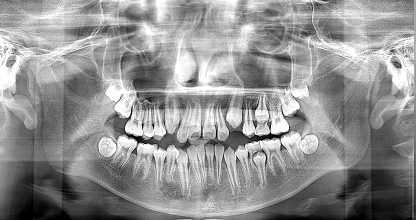
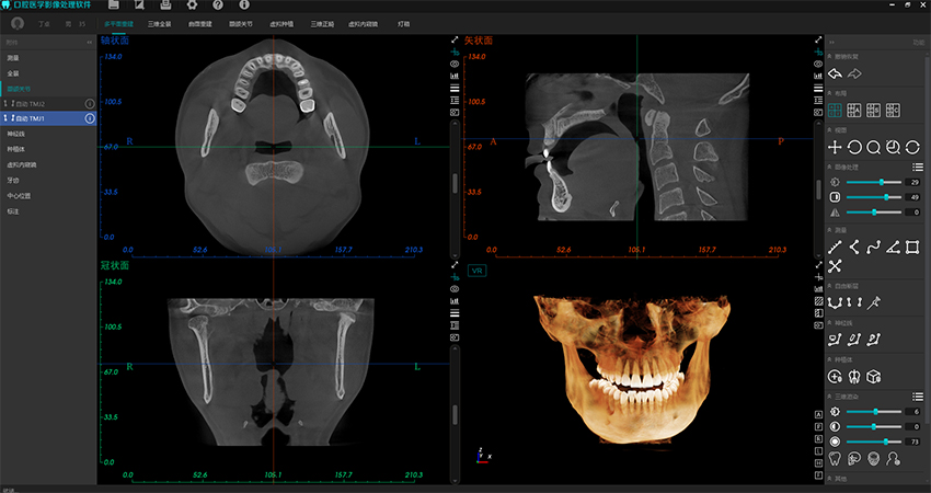
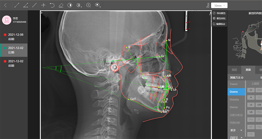
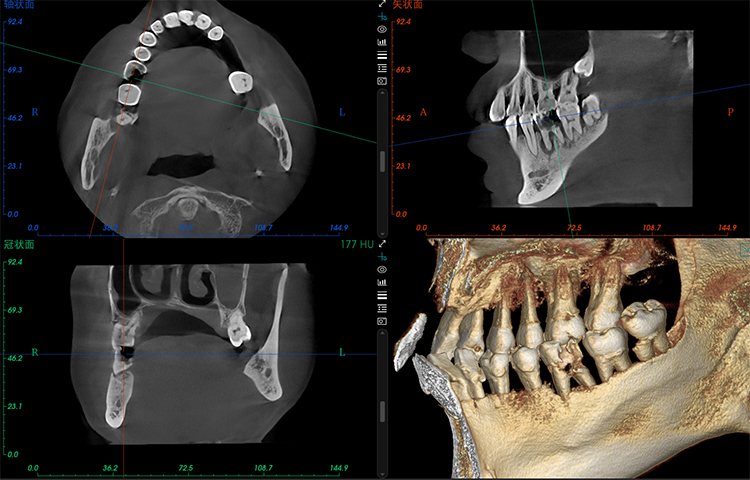
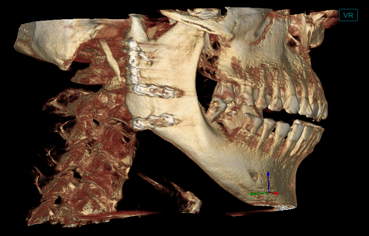
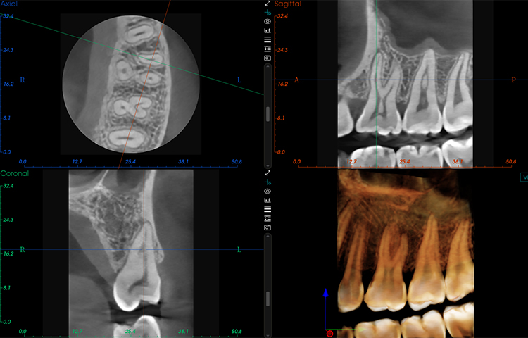
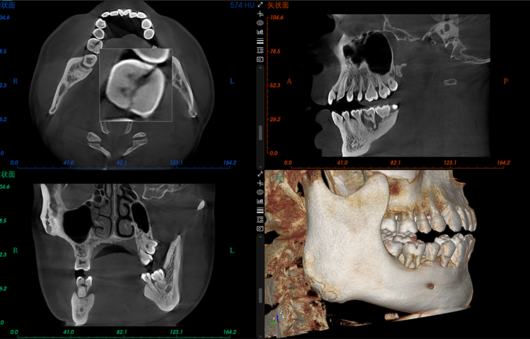
Clear imaging, meeting comprehensive clinical diagnosis and treatment needs
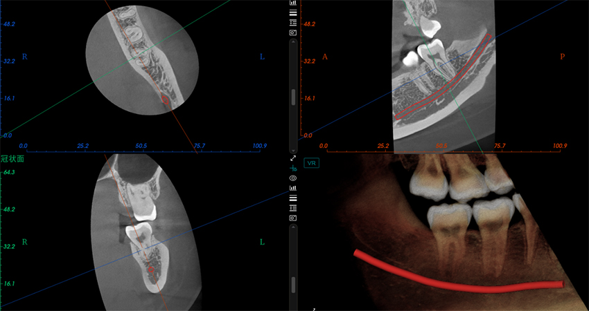
Rich functionality significantly enhances doctor-patient communication efficiency
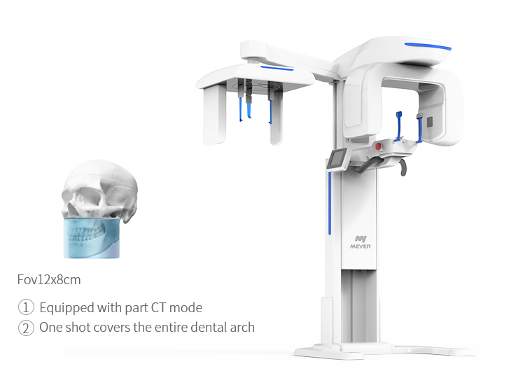
- Fov12×8cm
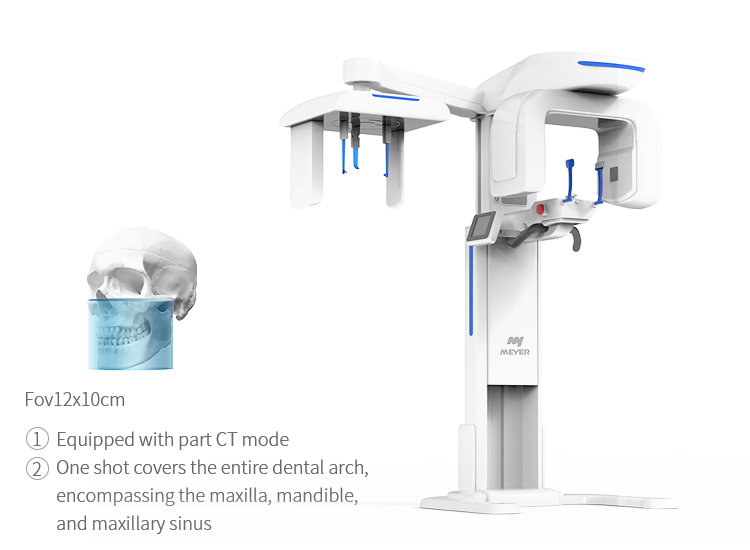
- Fov12×10cm
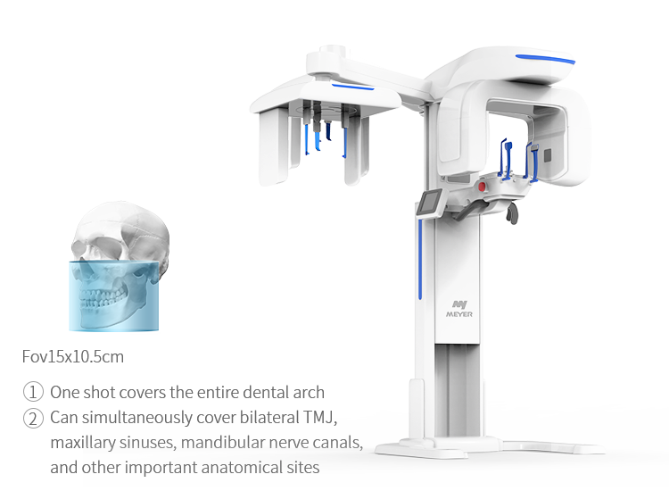
- Fov15×10.5cm
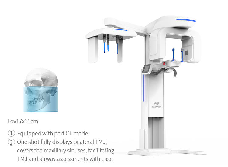
- Fov17×11cm
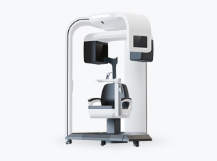
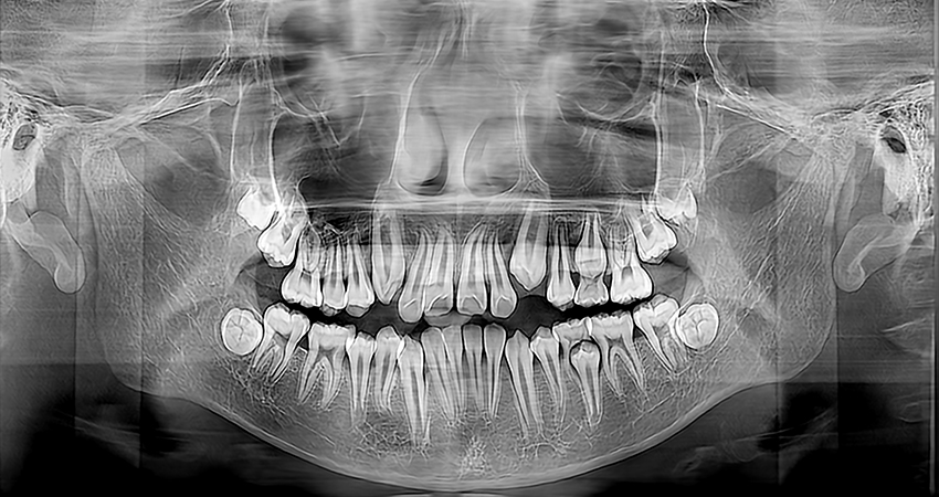
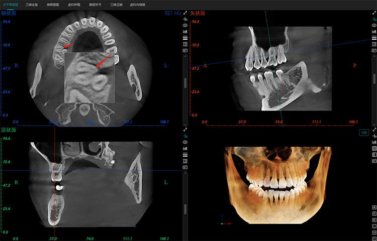
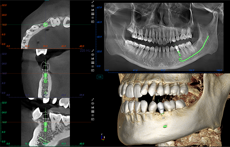
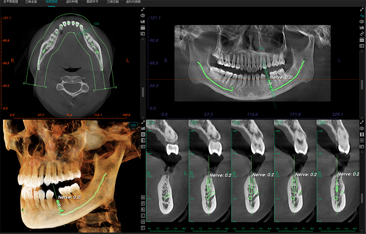
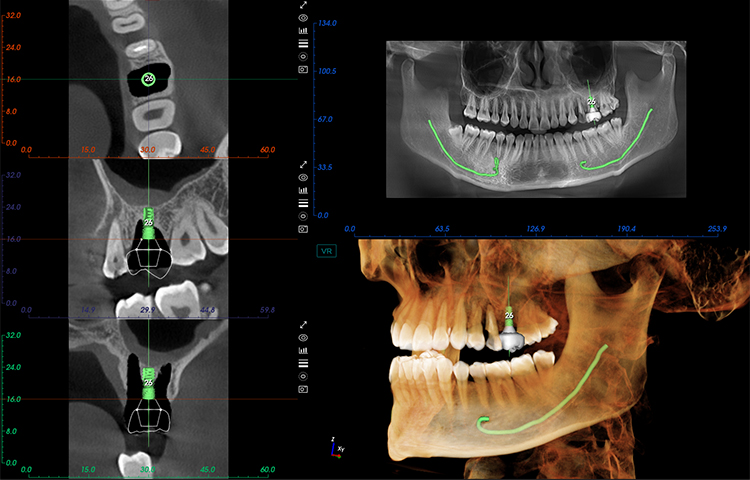
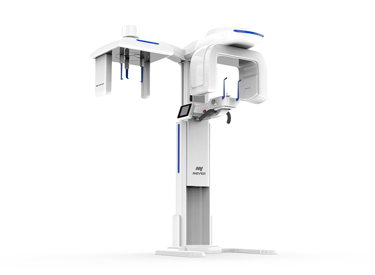
Meyer Dental CBCT is primarily utilized for comprehensive clinical diagnosis and treatment in dental implantology, orthodontics, restorative dentistry, oral and maxillofacial surgery, and endodontics.
A single capture can generate panoramic, CBCT, and Ceph images, providing an objective reflection of the 3D spatial structure of oral and maxillofacial tissues. This offers objective evidence for disease diagnosis, treatment planning, and prognosis assessment.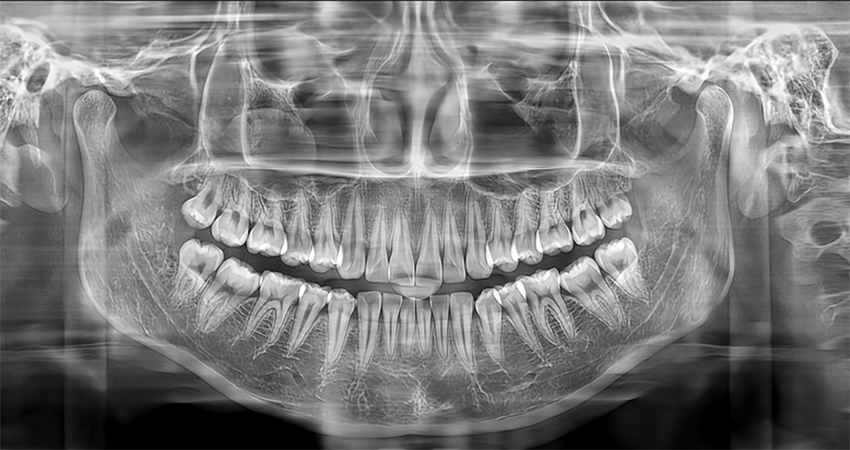


 Fov12×8cm
 Fov12×10cm
 Fov15×10.5cm
 Fov17×11cm
|
Capturing Mode |
CBCT/Pano/Ceph/Model Scan/Part CT/TMJ |
|
X-ray Tube Voltage |
60~90kv |
|
X-ray Tube Current |
2~10mA |
|
Image Detector |
CsI (Cesium Iodide) flat panel detector with large dynamic range |
|
Scan Time (Exposure Time) |
CBCT:20s(8.7s), Pano:17s Ceph:12s, Endo:12s |
|
Fov |
Maximum FOV (Field of View) in a single shot: 15x10.5cm |
|
Focal Spot |
0.5mm(IEC 60336) |
|
Reconstruction Time |
CT: < 60s |
|
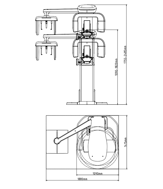
Weight |
220kg - 485lbs(without Ceph) 260kg - 573lbs(with Ceph) |
|
SS-X9010Dpro-3D |
CBCT/Pano/Model Scan/Part CT |
|
SS-X9010Dpro-3DE |
CBCT/Pano/Ceph/Model Scan/Part CT |
|
Capturing Mode |
CBCT/PANO/Ceph/Model Scan/Part CT/TMJ |
|
X-ray Tube Voltage |
60~90kV/60~100kV |
|
X-ray Tube Current |
2~10mA/4~10mA |
|
Image Detector |
CsI (Cesium Iodide) flat panel detector with large dynamic range |
|
Exposure Time |
CBCT 20s/16s,Pano 17s,Ceph 12s |
|
Focal Spot |
0.42, 0.31, 0.25, 0.18, 0.12, 0.07mm |
|
Fov |
FOV Maximum FOV (Field of View) in a single shot FOV:14x10 / 13x10cm |
|
3D Image Acquisition Technology |
Single-shot 360-degree Cone-beam Technology without Stitching |
|
Gray Scale |
CT: 16bit |
|
Reconstruction Time |
CT: < 60s |
|
SS-X9010Dpro-3D |
CBCT, Pano, Model Scan, Part CT |
|
SS-X9010Dpro-3DE |
CBCT, Pano, Ceph, Model Scan, Part CT |
|
SS-X10010DPlus |
CBCT, Ceph, Pano, Model Scan, Part CT, TMJ Scan |
|
Capturing Mode |
CBCT/Pano/Ceph/Model Scan/Part CT/TMJ |
|
X-ray Tube Voltage |
60~90kv |
|
X-ray Tube Current |
2~10mA |
|
Image Detector |
CsI (Cesium Iodide) flat panel detector with large dynamic range |
|
Exposure Time |
CBCT 20s/Pano 17s/Ceph 12s |
|
Fov |
Maximum FOV (Field of View) in a single shot: 15x10.5cm |
|
Focal Spot |
0.42/0.31/0.25/0.18/0.07mm |
|
Reconstruction Time |
CT: < 60s |
|
Weight |
CT: 265 kg |
|
SS-X9010Dpro-3D |
CBCT/Pano/Model Scan/Part CT |
|
SS-X9010Dpro-3DE |
CBCT/Pano/Ceph/Model Scan/Part CT |
|
Capturing Mode |
CBCT/Pano/Ceph/Model Scan/Part CT/TMJ |
|
X-ray Tube Voltage |
60~100kV |
|
X-ray Tube Current |
4~10mA |
|
Image Detector |
Amorphous Silicon Flat Panel Detector |
|
Exposure Time |
CBCT 16s, Pano 17s, Ceph 12s, TMJ 20s |
|
Focal Spot |
0.42, 0.31, 0.25, 0.18, 0.12, 0.07, 0.041mm |
|
Fov |
Maximum FOV (Field of View) in a single shot: 17x11cm |
|
3D Image Acquisition Technology |
Single-shot 360-degree Cone-beam Technology without Stitching |
|
Gray Scale |
CT: 16bit |
|
Reconstruction Time |
CT: < 60s |


-


-

 CONTACT ONLINE